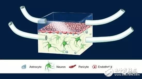
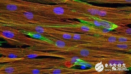
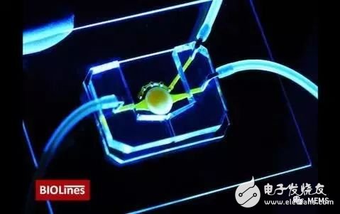
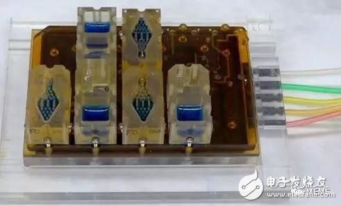
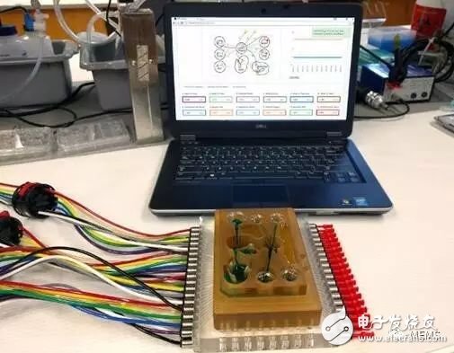
According to the Memes Consulting, from the beating heart to the breathing lung, organ chips have become one of the most popular emerging tools in human biology research. Although these devices may be closer to computer components than human parts, scientists have created working models for all organs, including the liver, lungs, and even the female reproductive system. Researchers hope to use these devices to simulate disease and promote drug development. Donald Ingber, director of Harvard University Wyss Institute for Biologically Inspired Engineering, told the magazine "scientists", "I think for most people, the common goal is a more efficient way to replace animal experiments and expand the personalized medicine." Small lung An alveolar chip with gas-filled (yellow) and medium-filled blood (blue) channels, lined with human cells to mimic organ function. According to Mamms Consulting, scientists at the Wyss Institute have developed 15 Different human organ chips. The first organ chip developed by the institute is a lung organ chip. It is a transparent, U disk-sized device with two channels. The air-filled upper channel is lined with alveolar epithelial cells, and the lower channel is lined with vascular cells, containing white blood cells. The solution will flow through the channel. To more closely mimic human biology, researchers used vacuum to deform the hollow tube in the main channel to simulate respiratory motion. “The research team’s innovation on the chip is based on my research, and the results show that mechanical forces are as important as chemistry and genetics for tissue development, maintenance, and function,†Ingber points out. “This alveolar chip can mimic normal organs. The physiological level and disease, the importance of discovering physical power, the search for new therapeutic targets, and even the development of new drugs, provide the original theory." Ingber and colleagues also introduced mechanical external forces into other organ chips, such as simulating intestinal peristalsis. A moving intestinal chip, as well as a kidney chip that simulates a pulse of blood vessels. Human bronchial epithelial image close-up on the respiratory tract chip, mucus transport cilia (pink) extending from the epithelial cells (cyan) to the air-filled lumen. One of the latest inventions of the research team is the airway chip, in addition to the replaced alveolar cells. The device is similar to the original lung chip, which is lined with human bronchial epithelial cells. The team used these chips to simulate chronic obstructive emphysema and asthma. They also use these devices to study the effects of smoking on bronchial epithelial cells, which are connected to a machine that ignites cigarettes and exhales smoke, similar to human smoking behavior. Creating a blood-brain barrier The illustration of the NeuroVascular Unit (NVU) chip, a human blood-brain barrier model. The porous membrane separates the brain modeling chamber and the chamber representing the peripheral vascular system. According to Memes Consulting, John Wikswo, a biomedical engineer from Vanderbilt University, and his team have created a A chip that studies the brain and blood-brain barrier. Wikswo writes told Scientists in an interview email, "We chose to study human neurovascular units because the interaction between cortical neurons and the blood-brain barrier (BBB) ​​that protects them is extremely important. Therefore, it is called a neurovascular unit. The human neurovascular unit chip consists of a microcavity that separates a chamber representing the brain and a chamber representing the peripheral vascular system through a porous membrane. The chip contains cortical neurons, microvascular endothelial cells, astrocytes, and perivascular cells from the human body. According to Wikswo, this organizational structure "allows us to study the metabolic responses of neurons and other cells to drugs and inflammatory signals that are transmitted through the blood-brain barrier." The team has incorporated neurovascular unit chips into a range of applications, such as investigating disease states and studying the effects of inflammation on disease. Currently, the team is also launching a new project to use this technology to help the pharmaceutical industry conduct drug testing. Beating heart Microscopic images of mouse heart tissue on the chip, cardiomyocytes (red), nuclei (blue), and actin (green) According to Memes Consulting, Megan McCain is a University of Southern California creature. A professor of medical engineering dedicated to the study of cardiac chips, which can hold live, beating heart cells in eraser-sized devices. The first step in creating the device is to extract the skin cells from the patient and reprogram them into stem cells, which are then cultured into cardiomyocytes. The second step is to place these cells on the surface of the chip made by bioengineering to create the natural environment of the heart. McCain pointed out that "the key indicator we are interested in is the generation of force." In 2014, McCain, who was also working at the Wyss Institute, used a cardiac chip to simulate Barth syndrome, a rare form of myocardium. Attenuate related genetic diseases. Currently, her team is working on using heart chips to study other diseases. McCain told the magazine "scientist", "Establishment of heart disease model is I think the chip can play the biggest role, in particular, is a genetic disease. Even if we give knockout mouse model, we can not capture all aspects of the human disease." Simulation eye An eye chip with a microfluidic channel (yellow) that carries nutrients to cells located in the center of the circular stent. The team also developed a micro-engineering eyelid that simulates a post-doctoral study on the chip that was once studied at Ingber Labs. Dan Huh, a bioengineering professor at the University of Pennsylvania, and his colleagues created this Eye chips, as well as eyelids that can blink. The chip is about the same size and shape as the contact lens, close to the size of the ocular surface. The chip contains human cells from the cornea and conjunctiva (covering the mucosal layer of the eye). The research team also designed the eyelids, which are attached to the surface of the chip to simulate the effect of blinking, helping to keep the surface of the chip lubricated. Huh said, "We found that blink movement is critical to the maintenance of the surface tissue of the eye. We are using the platform to simulate certain chronic eye diseases, such as dry eye." According to Huh, his experimental team plans to use eye chips. Simulate other eye conditions for drug testing and development, as well as further testing and optimizing contact lenses. The team is also currently developing retina chips. “Eye is one of the key areas of research in our laboratory.†Huh emphasized that he and his team also studied other types of organ chips, including the lungs and placenta. Simulated menstruation EVATAR is a pocket-sized female reproductive system model. The blood sample fluid (blue) will flow through the pores containing micro-organs. According to Memes Consulting, many research teams want to integrate different organ chips to recreate organs. System, even the entire human body model. Teresa Woodruff, a professor of obstetrics and gynaecology at Northwestern University, and colleagues integrated five miniature organs on a palm-sized chip and successfully established a model of the female reproductive tract. The chip, called EVATAR, consists of a series of tubes and micropumps. The blue blood sample flowing through the cells contains five mini organs: the fallopian tubes, the uterus, the vagina, the ovaries, and the liver. “The system helps us deliver media in a way that carries fresh nutrients and removes waste,†Woodruff said. “This way of operation is no different from the human body.†By adding hormones to the circulating fluid, the team was able to simulate a 28-day menstrual cycle. cycle. The research team hopes to use EVAAR to clarify reproductive physiology and related diseases, as well as related drug testing and development. They are also working on a male version of the chip and named it ADATAR. interconnected The researchers connected multiple organ platforms to related software, each connected to a micro-physiological system, such as the lungs, intestines, or central nervous system, through which fluids flowed to simulate physiological cardiac output. Linda Griffith, a professor of bioengineering at the Massachusetts Institute of Technology (MIT), and colleagues are one of two teams funded by the US Government's Defense Advanced Research Projects Agency (DAPRA), who created the "body chip." Connect 10 different mini organ systems to the integrated circuit. Another group of teams came from the Wyss Institute. According to Griffith's presentation, his team recently "completed an important milestone in DARPA, connecting 10 organ microphysiological systems together in a month." Connecting multiple organ chips can help researchers understand between organs. interaction. In their latest trial, Griffith and his team studied the role of inflammation in interconnected human intestinal chips and liver chips. This study shows how crosstalk between two organs affects gene expression and tissue-specific function, among other factors. Griffith said, "I think the field is currently in the early stages of complex physiology that considers the interaction of multiple cell types within organs and devices, especially when it comes to the interaction of tissue cells with the immune system. Our project is very large. Part of the focus is on the field of immunology." The artistic performance of human body chips is close to in vitro biology, and bioengineering equipment has trained many brain microarrays in space that can represent the smallest functional unit of each organ. Emulate's organ chips, such as brain chips, contain tiny hollow channels that line up thousands of living human cells and tissues, each with about AA batteries (5th battery). According to Memes Consulting, in order to study Wyss The organ chip technology developed was commercialized, and the startup Emulate was born. The company's products include pulmonary heart, liver chips and intestinal chips. Although the above chips have different cell types and functions, the standardized design makes them look similar. The company recently announced plans to send its brain chips to the International Space Station to study how, among other things, the blood-brain barrier, stress and inflammation affect brain function. The company's brain chips contain neurons and vascular endothelial cells that can be used to mimic physiological and blood-brain barriers. Geraldine Hamilton, general manager and chief scientific officer of Emulate, explains, “We are not just rebuilding the entire brain model, but also the smallest functional unit of the organ. In the above scenario, we take microvascular endothelial cells, neurons, and star glue. The blood-brain barrier of cytoplasmic and pericytes, which interact in a specific way and need to be organized in a specific way, which is what we need to recreate in the brain chip - such a small size." Portable Speaker,Speaker Receiver,Outdoor Speakers,Wireless Speakers GUANGZHOU LIWEI ELECTRONICS CO.,LTD , https://www.gdliwei.com